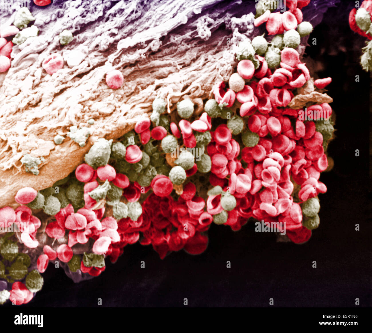
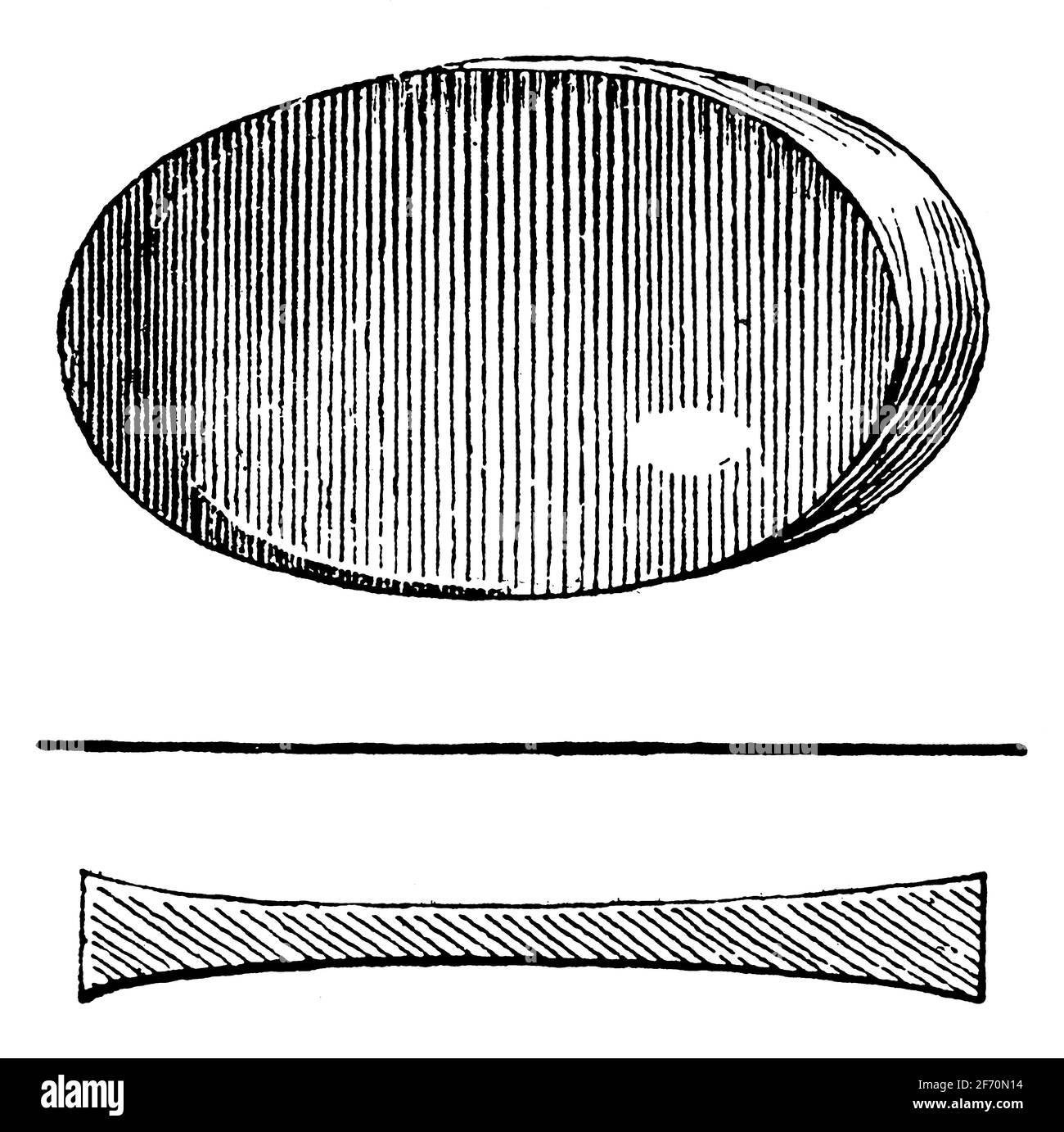
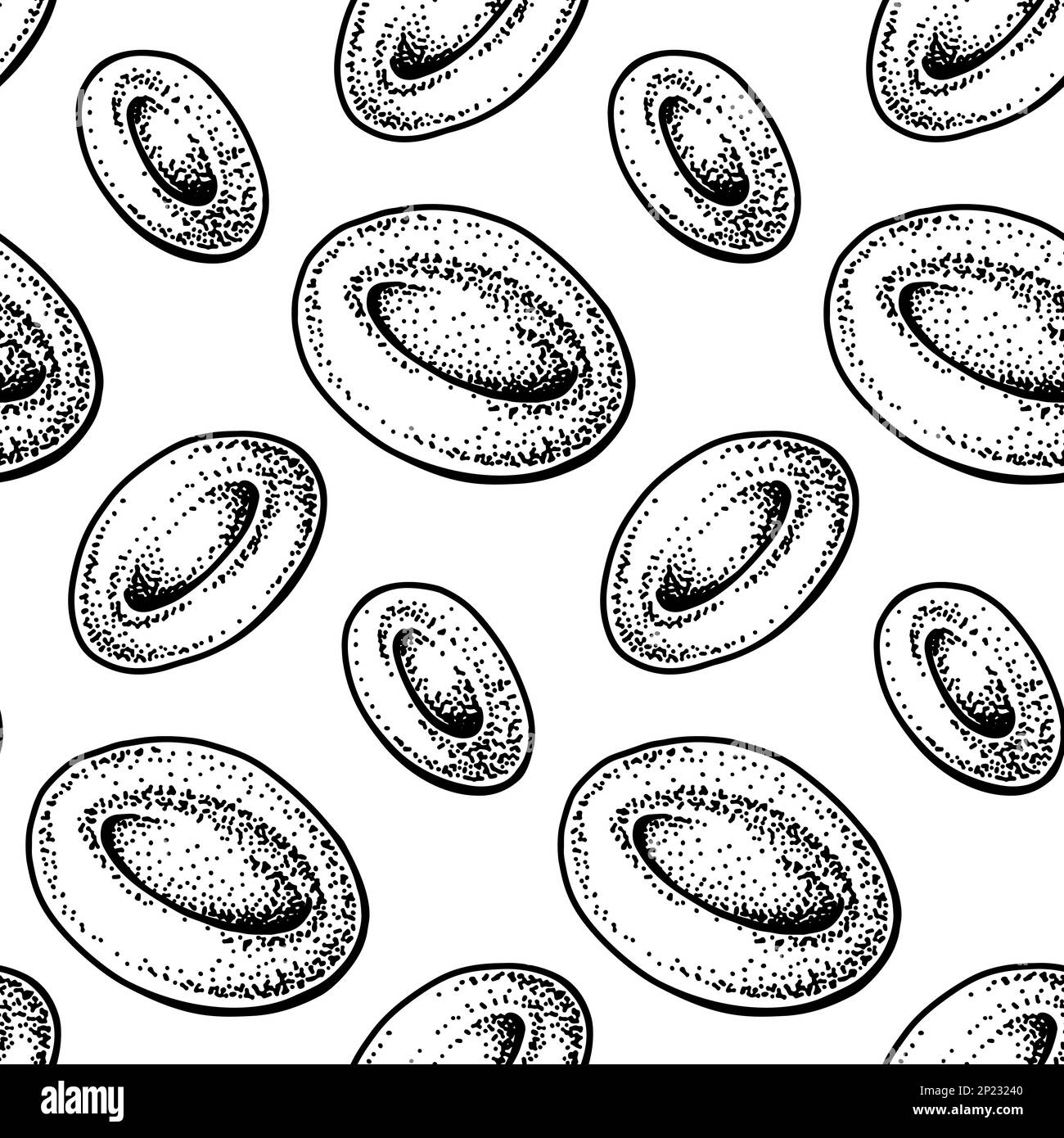
This scanning electron micrograph (SEM) depicted a number of red blood cells found enmeshed in a fibrinous matrix on the luminal surface of an indwelling vascular catheter; Magnified 11432x Note the biconcave
$ 18.00 · 4.5 (771) · In stock

Download this stock image: This scanning electron micrograph (SEM) depicted a number of red blood cells found enmeshed in a fibrinous matrix on the luminal surface of an indwelling vascular catheter; Magnified 11432x Note the biconcave cytomorphologic shape of each erythrocyte, which increases the surface area of these hemoglobin-filled cells, thereby, promoting a greater degree of gas exchange, which is their primary function in an in vivo setting. In their adult phase, these cells possess no nucleus. What appears to be irregularly-shaped chunks of debris, are actually fibrin clumps, which when inside the living organi - 2BE0H0B from Alamy's library of millions of high resolution stock photos, illustrations and vectors.
Fibrin red blood cells hi-res stock photography and images - Alamy
Scanning Electron Microscope Image of Blood Cells: Image Details - NCI Visuals Online
Red blood corpuscles hi-res stock photography and images - Alamy

RED CELL & FIBRIN
This scanning electron micrograph (SEM) depicted a closer view of number of red, Stock Photo, Picture And Rights Managed Image. Pic. BSI-1421505

Blood cells sem hi-res stock photography and images - Alamy
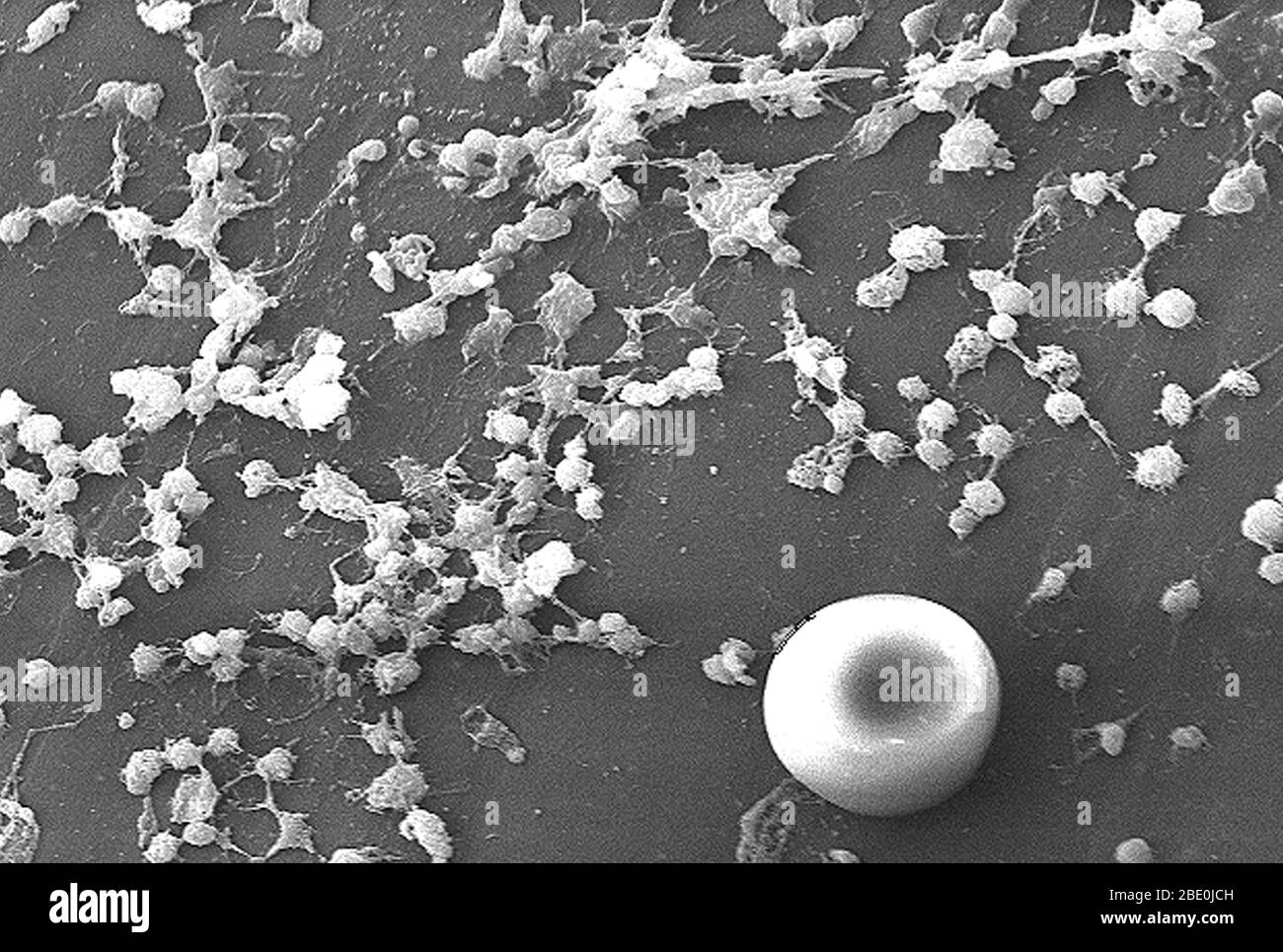
Scanning Electron Micrograph (SEM) depicting large numbers of Staphylococcus aureus bacteria, which were found on the luminal surface of an indwelling catheter. A red blood cell (RBD), also known as an erythrocyte
Red blood corpuscles hi-res stock photography and images - Alamy
Biconcave hi-res stock photography and images - Alamy
Red blood cells Black and White Stock Photos & Images - Alamy
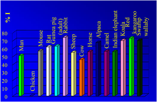
Frontiers Light and Scanning Electron Microscopy of Red Blood Cells From Humans and Animal Species Providing Insights into Molecular Cell Biology
![]()
Red blood corpuscles Black and White Stock Photos & Images - Alamy
Irregularly shape hi-res stock photography and images - Alamy
This scanning electron micrograph (SEM) depicted a number of red blood cells found enmeshed in a fibrinous matrix on the luminal surface of an indwelling vascular catheter; Magnified 7766x. In this instance