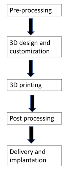
The STL images of two geometries of the 3D-printed bioceramic model
$ 21.99 · 4.5 (247) · In stock

Download scientific diagram | The STL images of two geometries of the 3D-printed bioceramic model were designed as follows: The cylindrical compression sample (a), the concave-topped disk structures views of the bottom (c), and the top (d). The cross-section views of concave-top disk structures also showed the STL image of a horizontal section (e) and a vertical section (f). Furthermore, the two kinds of 3D-printed sintered bioceramic images were obtained. The 3D cylinder bioceramic sample (b), the bottom view (g), and the top view (h) of the concave-top disc structure of the 3D-printed bioceramic scaffold from publication: Bilayer osteochondral graft in rabbit xenogeneic transplantation model comprising sintered 3D-printed bioceramic and human adipose-derived stem cells laden biohydrogel | Reconstruction of severe osteochondral defects in articular cartilage and subchondral trabecular bone remains a challenging problem. The well-integrated bilayer osteochondral graft design expects to be guided the chondrogenic and osteogenic differentiation for stem cells and | Bioceramics, Osteochondritis and Grafts | ResearchGate, the professional network for scientists.
IJMS, Free Full-Text

Challenges on optimization of 3D-printed bone scaffolds

Bioengineering, Free Full-Text

5776 PDFs Review articles in NANO-SILICA

Three-dimensional printing of biomaterials for bone tissue

The thermogravimetric (TGA) analysis of the 3D-printed bioceramic
A) Top and crosssectional confocal images of the actin filaments

PDF) Vascularized Bone Tissue Engineering: Approaches for

Verifying Structural Integrity of Metal 3D-Printed Parts

3D modeling of the calcaneus and 3D printed models. (a) The
Make 3d printing by wattronix - Issuu

Che Wei WU, Doctor of Philosophy

The STL images of two geometries of the 3D-printed bioceramic